
Body Composition

Nicholas A. Buoncristiani, MS
Doctoral Student
University of North Carolina at Chapel Hill
Chapel Hill, North Carolina, United States
James Merritt
R&D Scientist
Gatorade Sports Science Institude
Valhalla, New York, United States
Evangeline P. Soucie
Clinical Research Associate
University of North Carolina Rex Cancer Center
Raleigh, North Carolina, United States
Hayden K. Giuliani-Dewig
Research Scientist
Rockefeller Neuroscience Institute at West Virginia University
Morgantown, West Virginia, United States
 photo")
Gena R. Gerstner, PhD, MPH, CSCS (she/her/hers)
Research Assistant Professor
University of North Carolina at Chapel Hill
Chapel Hill, North Carolina, United States
Abbie E. Smith-Ryan
Professor
University of North Carolina at Chapel Hill
Chapel Hill, North Carolina, United States
Brian Pietrosimone
Professor
University of North Carolina at Chapel Hill
Chapel Hill, North Carolina, United States
Eric D. Ryan
Professor
University of North Carolina at Chapel Hill
Chapel Hill, North Carolina, United States
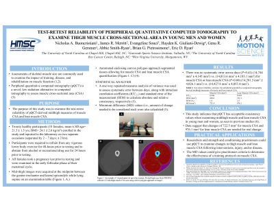
Assessments of skeletal muscle size are commonly used to examine the impact of training, disease, and rehabilitation on muscle function. Peripheral quantitative computed tomography (pQCT) is a novel, low-radiation alternative to computed tomography to assess muscle cross-sectional area (CSA).
Purpose: The purpose of this study was to examine the test-retest reliability of pQCT-derived midthigh measures of muscle CSA and lean muscle CSA.
Methods: Twenty healthy participants (10 females; mean ± SD age= 21.3 ± 1.5 yrs, BMI= 24.1 ± 2.6 kg/m2) enrolled in the study and reported to the laboratory on two separate occasions (separated by 2 – 7 days) at the same time of day (± 2 hrs). Prior to both sessions, participants were required to refrain from any vigorous lower body exercise or resistance training for 48 hours, and not engage in any form of exercise and abstain from alcohol or recreational drug use for 24 hours prior to testing. All females took a pregnancy test prior to testing and were examined in the early follicular phase of their menstrual cycle for both testing sessions. Mid-thigh images were acquired at the midpoint between the greater trochanter and lateral epicondyle while lying supine on an examination table. Freely available software was used to determine muscle and lean muscle (excluding non-contractile tissue) mid-thigh CSA assessments. A one-way repeated measures analysis of variance was used to assess for systematic error between days. The intraclass correlation coefficient (ICC2,1) and standard error of the measurement (SEM) were calculated to assess relative and absolute consistency, respectively. Minimum difference (MD) values (i.e., amount of change needed to be considered real) were also calculated.
Results: Test-retest reliability statistics are reported in Table 1. There was no systematic error between day one (14,744.4 mm2 ± 4,145.0 mm2) and day two (14,813.6 mm2 ± 4,183.1 mm2) for muscle CSA (P= 0.41) or lean muscle CSA (P = 0.60) between day one (14,281.5 mm2 ± 4,026.1 mm2) and day 2 (14,427.8 mm2 ± 4,053.4 mm2).
Conclusions: Results from this study indicated that pQCT has acceptable relative and absolute consistency values when examining midthigh muscle and lean muscle CSA in young men and women. These data suggest that changes of 722.5 mm2 for muscle CSA and 976.1 mm2 for lean muscle CSA would be needed to be considered a real change.
PRACTICAL APPLICATION: Researchers and strength and conditioning practitioners could consider using pQCT to reliably examine changes in thigh muscle and lean muscle CSA following various interventions, injury, and/or chronic disease. Furthermore, the MD values could give practitioners specific criteria to determine the effectiveness of a training or rehabilitation protocol on muscle CSA.