
Biomechanics/Neuromuscular

McKenzie M. Hare
Graduate Research Assistant
Texas Tech University
Northville, Michigan, United States

Kealey J. Wohlgemuth, MA, CSCS,*D, CISSN
Graduate Part-Time Instructor
Texas Tech University
Surf City, North Carolina, United States

Kathryn E. Southall, BS
Graduate Research Assistant
Texas Tech University
Englewood, Colorado, United States
.jpg "Malia N.M Blue, MA photo")
Malia N.M Blue, MA
Assistant Professor
University of North Carolina at Chapel Hill
Chapel Hill, North Carolina, United States

Katie G. Kennedy, MS
Graduate Assistant
University of South Alabama
Mobile, Alabama, United States
Jacob A. Mota
Assistant Professor of Kinesiology
Texas Tech University
Lubbock, Texas, United States
Muscle anatomical cross-sectional area (ACSA) and echo intensity (EI) are commonly assessed with brightness-mode (B-mode) ultrasound. Scans taken using B-mode ultrasound are manually analyzed by an experienced technician and require consistent selection of muscle tissue, while avoiding the surrounding fascia, making the task extremely time-consuming. Furthermore, utilizing a manual analysis technique may result in an increased error of the measures due to technician bias. Recently, an automatic software was created to eliminate technician bias and decrease the time required for ultrasound analysis.
Purpose: The purpose of this study is to investigate the validity of manual compared to automatic ultrasound analysis techniques for muscle anatomical cross-sectional area and echo intensity of the vastus lateralis.
Methods: Twenty-two individuals (mean ± SD; Age = 24 ± 4 yrs; BMI = 24.19 ± 3.26 kg/m2) volunteered for this study. Participants completed a single data collection trial consisting of panoramic ultrasound images acquired at frequencies of both 10 MHz and 12 MHz. B-mode ultrasound settings were held constant with depth = 6 cm and gain = 52 dB, and scans were taken of the vastus lateralis (VL) at 50% the length of the proximal to distal musculo-tendon junctions. Ultrasound images were analyzed manually by an experienced technician with open-source image analysis software. To determine ACSA, images were traced using the polygon tool, localizing the muscle and area of interest. To quantify EI, the histogram function was used on the same traced area of muscle. Automatic analysis was completed with the Deep Anatomical Cross-Sectional Area (DeepACSA) program. DeepACSA is an algorithm programed to automatically trace the area of interest on an ultrasound image. The program automatically calculates the ASCA and EI for each ultrasound image. Validity statistics (i.e., constant error [CE], total error [TE], and standard error of the estimate [SEE]) and Bland-Altman plots were calculated for both the manual and automatic analyses for each ultrasound frequency.
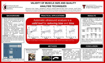
Results: When validated against manual analyses, automatic analyses of ACSA presented greater TE and SEE at a frequency of 10 MHz (CE = -0.73 cm2, TE = 4.77 cm2, SEE = 3.56 cm2) than at 12 MHz (CE = 0.16 cm2, TE = 3.29 cm2, SEE = 3.03 cm2). Analyses of EI presented a greater TE and SEE at a frequency of 10 MHz (CE = 3.21 au, TE = 4.17 au, SEE = 2.52 au) than at 12 MHz (CE = 3.07 au, TE = 4.01 au, SEE = 2.29 au). The regression lines in the Bland-Altman plots indicate a bias for analysis of ACSA (10 MHz: -0.76, 95% confidence interval [CI] = -2.95 to 1.43, R2 = 0.726; 12 MHz: 0.17, CI = -1.36 to 1.69, R2 = 0.778) and for analysis of EI (10 MHz: 3.35, CI = 2.15 to 4.55, R2 = 0.910; 12 MHz: 3.21, CI = 2.05 to 4.37, R2 = 0.905).
Conclusions: Automatic ultrasound analyses of the VL were validated against manual ultrasound analyses. The results of this study demonstrated the automatic program had low CE, TE, and SEE, regardless of frequency. This suggests DeepACSA may be a promising program. However, it is encouraged investigators use caution when utilizing the program until further investigation is completed. PRACTICAL APPLICATION: These findings may be useful for the investigation of skeletal muscle size and quality. Investigators may utilize this automatic program for ultrasound analyses to decrease the time and technician bias accompanied by manual analysis.