
Special Populations

Cory M. Smith, PhD
Assistant Professor
Baylor University
Waco, Texas, United States

Cierra Ugale, MS
PhD Student / Research Assistant
Baylor University
Waco, Texas, United States
Kate Lee
Graduate Research Assistant
Baylor University
Waco, Texas, United States
Andrew R. Gallucci
Associate Professor
Baylor University
Waco, Texas, United States

Matt Segovia, MS
PhD Student / Research Assistant
Baylor University
Waco, Texas, United States
.jpg "Owen F. Salmon, MS photo")
Owen F. Salmon, MS
PhD Student / Research Assistant
Baylor University
Waco, Texas, United States
Purpose: Utilize functional near-infrared spectroscopy (fNIRS) derived total hemoglobin content (tHb) to identify the impact of a concussion while performing a 60s psychomotor vigilance test (PVT) in an athlete who received a direct blow to the temple.
Methods: A female collegiate athlete sustained a direct impact to the side of the temple from a collision during competition. The athlete was diagnosed with a concussion after evaluations completed by the team’s physician and athletic trainer. To assess the effect of the impact, 4 optode x 1 receiver arrangements were orientated into squares and placed over the injured (impacted) and uninjured temple to simultaneously compare ΔtHb during the PVT. The PVT lasted for 60s and assessed reaction time by tapping numbers on the center of a tablet as quickly as possible. The PVT was chosen as it requires the combined use of visual ques, neurocognitive loading, and physical movement. The fNIRS data for the injured and uninjured temple were used to calculate tHb which reflects overall blood flow to that region of the brain. This case study aimed to determine if a PVT coupled with fNIRS was capable of identifying tHb differences in regional brain health in a person with a concussion. A Morlet wavelet analysis was utilized to examine changes over 10s averages during the 60s PVT test for a total of 6 timepoints. Specific details of the athlete that may be identifiable have been excluded to maintain anonymity per IRB.
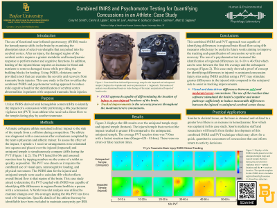
Results: Figure 1 displays the tHb results over the uninjured temple (top) and injured temple (bottom). The injured temple that received the impact resulted in greater tHb compared to the unimpacted. uninjured temple. The average PVT reaction time was 710ms with a fastest reaction time (Range=624-814ms). There were no errors or false reaction times.
Conclusion: This combined fNIRS and PVT approach was capable of identifying differences in regional brain blood flow using tHb measures which may be useful in future works aiming to improve the tracking and quantification of concussion severity and recovery. The use of a psychomotor test increased the identification of regional differences (ie. 0-10 vs 40-50s) which can be seen between the first 10s average and the subsequent averages (Figure 1). This case studied showed a proof of concept for identifying differences in injured vs uninjured concussion injury sites using fNIRS and that using a PVT may stimulate greater differences in the injured side compared to the uninjured side to assist in tracking improvements.
PRACTICAL APPLICATIONS: Similar to skeletal tissue, as the brain is strained and utilized to a greater level there is an increase in hemodynamic flow which was captured in this case study. Athletic trainers, team physicians, and researchers will benefit from further development of this combined fNIRS and PVT technique which may allow for a quick, quantitative assessment of concussions that can inform return to activity decisions.
Acknowledgements: None