
Biomechanics/Neuromuscular

Cierra Ugale, MS
PhD Student / Research Assistant
Baylor University
Waco, Texas, United States
.jpg "Owen F. Salmon, MS photo")
Owen F. Salmon, MS
PhD Student / Research Assistant
Baylor University
Waco, Texas, United States

Matt Segovia, MS
PhD Student / Research Assistant
Baylor University
Waco, Texas, United States

Cory M. Smith, PhD
Assistant Professor
Baylor University
Waco, Texas, United States
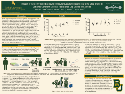
Purpose: Examine the influence of acute exposure to normobaric hypoxia on electromyographic (EMG) root mean square (RMS) and mean power frequency (MPF) on the vastus lateralis (VL) during leg extensions performed at various step intensities.
Methods: 13 recreationally active men (age: 21.2 ± 2.9 yr., height: 174.8 ± 12.8 cm, weight: 83.2 ± 14.1 kg) were exposed to two simulated altitude conditions, normoxic (N; Fraction of inspired oxygen [FiO2] = 21%) and a hypoxic condition (H13; FiO2 = 13%, simulating ~12,500 ft.). Each participant visited the laboratory three times separated by 24-48 hours. Visit 1 was a familiarization visit to establish a 1 repetition maximum (1RM) leg extension and determine load intensities for the step muscle actions that will be used for each condition. During the two testing visits, the participants were randomly assigned to either the N or H13 conditions and performed step muscle actions unilaterally in a randomized order at 20%, 40%, 60%, 80% and 100% of their 1RM leg extension following a proper warm-up. During each leg extension, EMG was recorded from the participants right VL, and was further analyzed to assess changes in the RMS and MPF during the concentric phase of each repetition. Two separate, 2 (Condition: N and H13) x 5 (step muscle actions: 20%, 40%, 60%, 80% and 100% of their 1RM leg extension) two-way repeated measure ANOVA was performed on EMG RMS and EMG MPF normalized to their N 1RM.
Results: For EMG RMS, there was no significant interaction (p = 0.37) or main effect for Condition (p = 0.74). There was a main effect for Intensity (p < 0.01) which indicated that 20% < 40% = 60% < 80% < 100% 1RM (p < 0.01 - 0.03). Means and standard deviation for EMG RMS at each step muscle actions were (mean±SEM: 20% = 61.9 ± 4.7, 40% = 79.3 ± 5.6, 60% = 80.2 ± 5.5, 80% = 86.2 ± 6.9, 100% = 94.5 ± 5.1% 1RM). For EMG MPF, there was no significant interaction (p = 0.83) or main effect for Condition (p = 0.17). There was a main effect for Intensity (p = 0.02) which indicated that 20% = 40% = 60% < 80% < 100% (p < 0.01). Means and standard deviation for EMG RMS at each step muscle actions were (mean±SEM: 20% = 107.2 ± 5.1, 40% = 115.4 ± 5.1, 60% = 117.9 ± 4.8, 80% = 118 ± 4.5, 100% = 118.8 ± 2.8% 1RM).
Conclusion: These results indicate that acute hypoxic exposure did not affect the neuromuscular responses during step muscle actions. However, there was an increase with each intensity for EMG RMS, except between 40% - 60 % 1RM, suggesting an increase in muscle activation with an increase in intensity. EMG MPF remained similar from 20% - 80% and then increased at 100% 1RM suggesting an increase in neuromuscular excitation at maximal intensities. PRACTICAL APPLICATIONS: These findings are useful for researchers and certified strength & conditioning specialists who are looking for methodologies to track the impact that intensity and acute hypoxia has on an athletes’ performance. Specifically, that neuromuscular responses track increases in intensity, but may not be ideal for evaluating the impact of hypoxia on muscular performance in an unfatigued state.
Acknowledgements: None